

In this Section...
Methods Applied to Orsten Fossils: From 2D to 3D Reconstructions and further to 3D Modelling and 3D Printing
4.1 Start with drawing and still working fine
Already Klaus Müller started his work on the 'Orsten' fossils with drawings of the first discoveries be made by non-scientific assistants (right: 1st reconstruction and later one, drawn by DW).
Subsequently, Dieter Waloszek took over this job and provided the reconstructions for Klaus' 1982 paper on Hesslandona unisulcata and the 1983 paper on six of the crustaceans in the material. Step by step he advanced the drawing techniques, re-drew photographs using transparency paper, overlaid images and combined photographs, scanned images and drawings to create 2D and shaded graphics of overviews and details to generalized views.
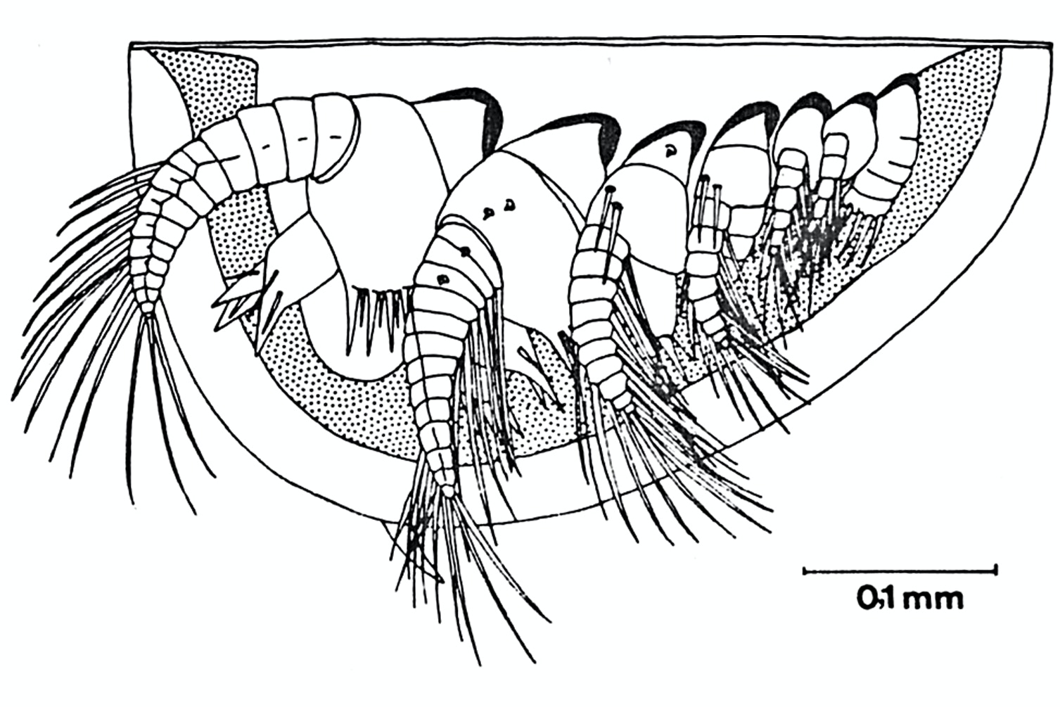
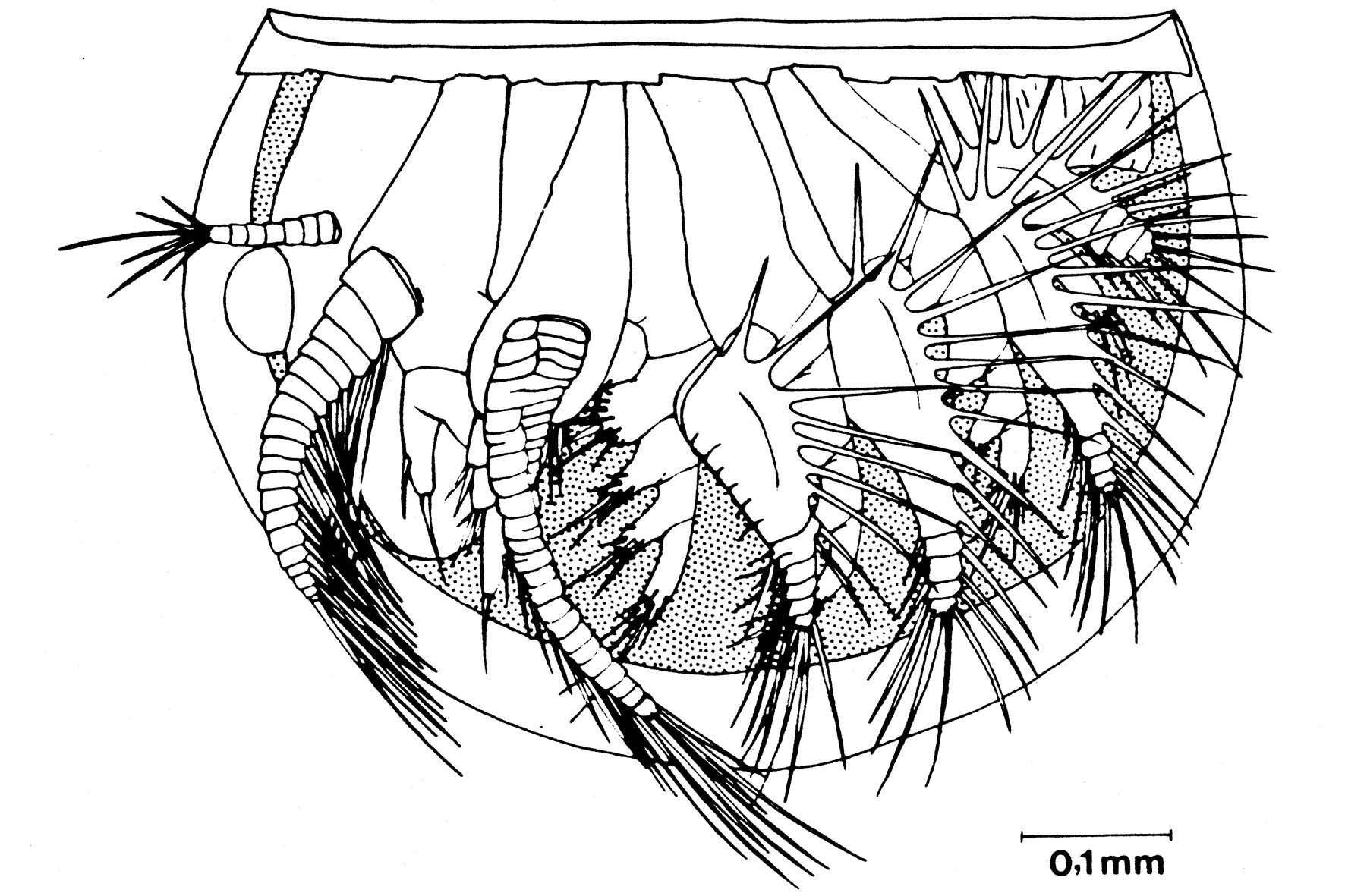
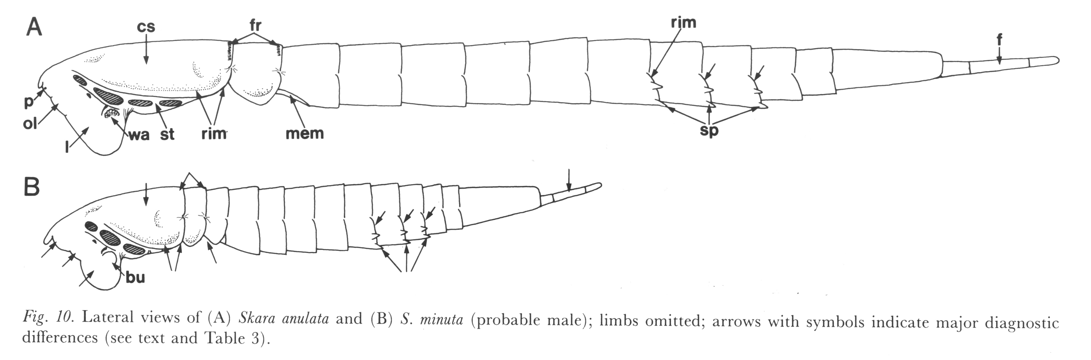
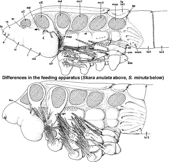
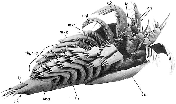
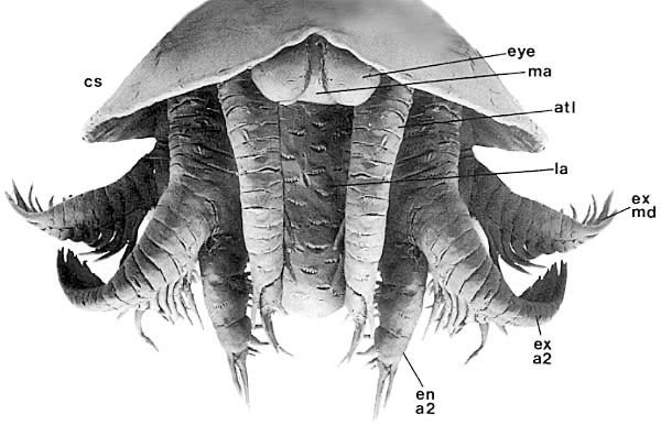
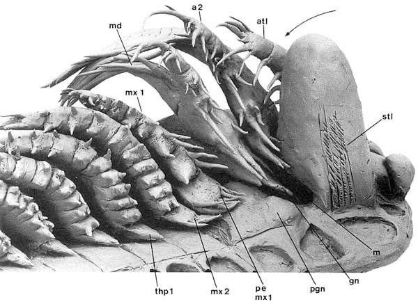
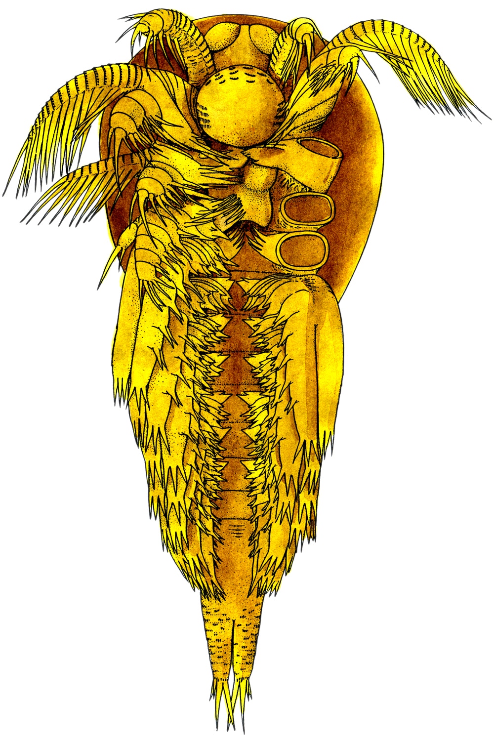
Examples are given here from the 1985 monograph on Skara ("Skaracarida"), a pioneering work providing all major techniques for the later studies. The left illustration shows the two species of Skara found in the 'Orsten' material, apparently co-existing: S. anulata, 1.2 mm long, and S. minuta, just 0.7 mm long. The smaller species differs from the larger species in e.g. a half as long head and trunk with segments, a different furca, and appendages with much finer and denser setation. Furthermore, the mandibular coxa ends in S. anulata in two spines, while the smaller species has a grinding plate with fine tooth-like cutting edge.
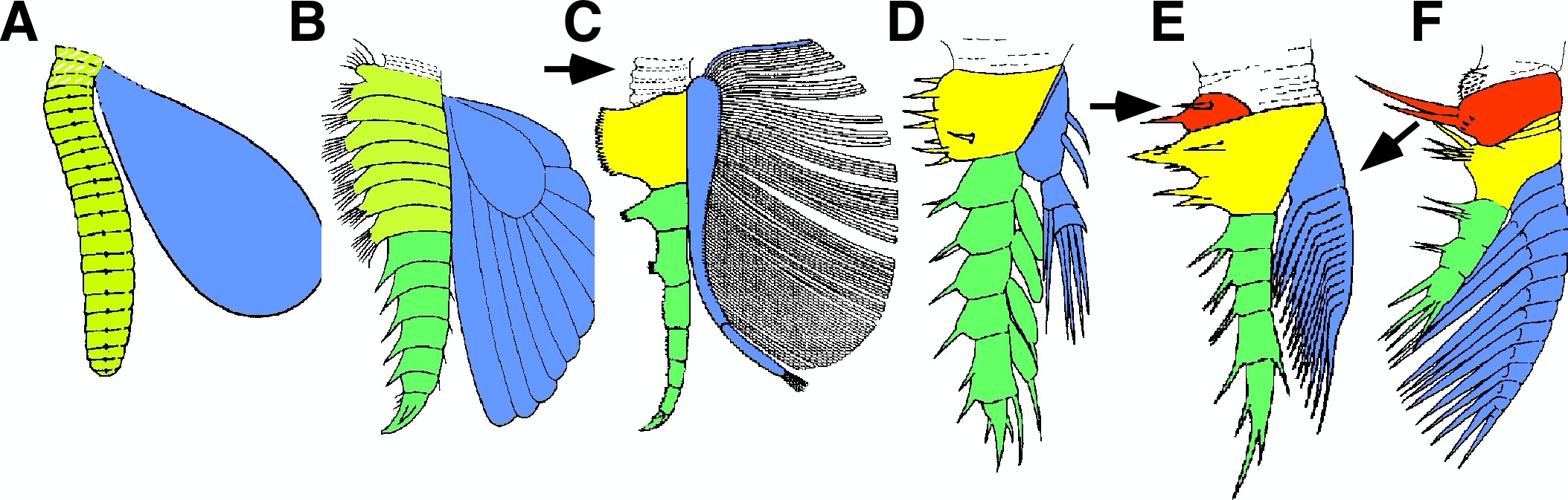
Later, we could also publish colour drawings with, which remained, however, an expensive adventure – meaning: a rare event. Therefore, we used coloured images of our material mainly for presentations at conferences or on our website. Here a colour scheme to put forward our ideas on the evolution of arthropod appendages.
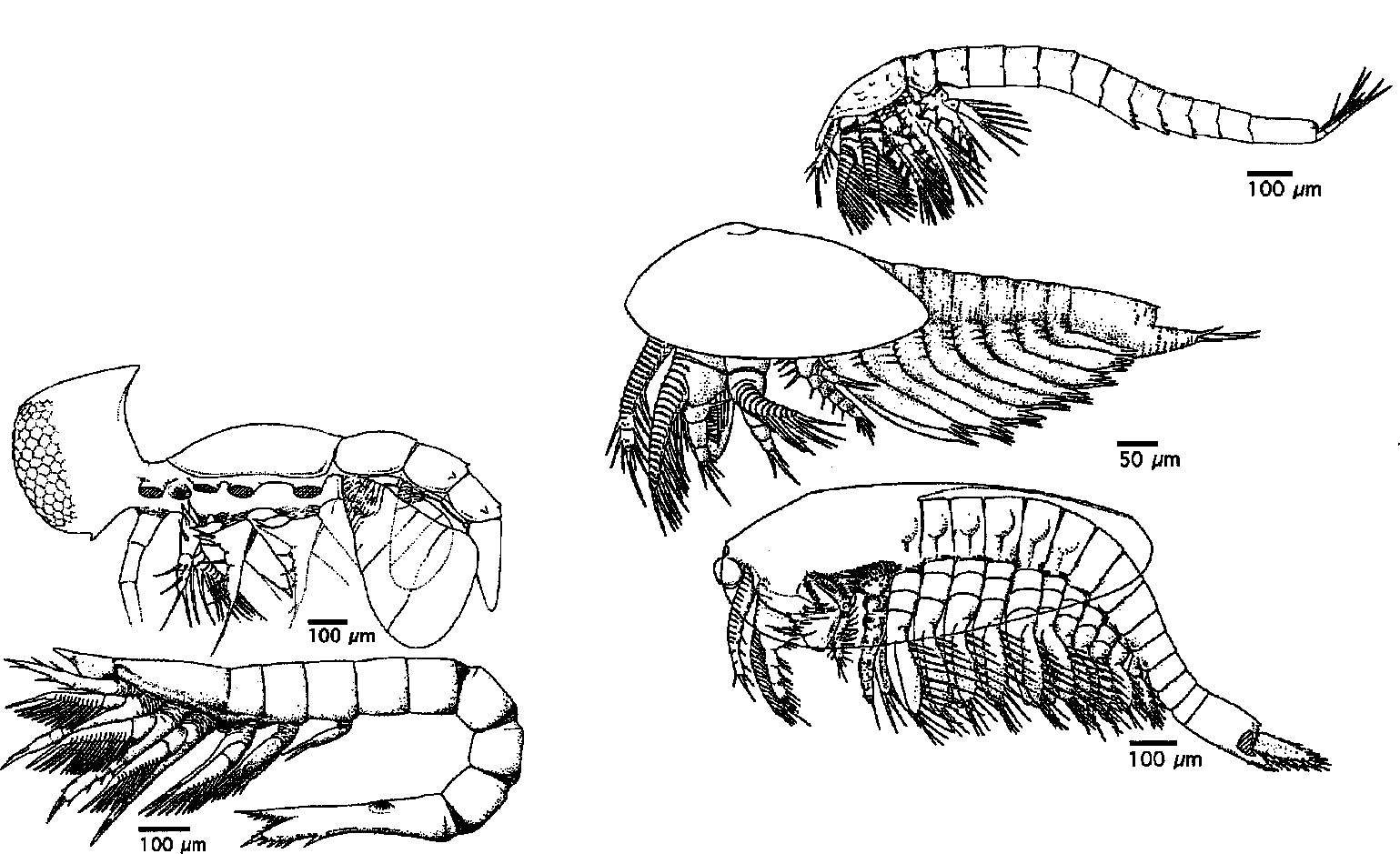
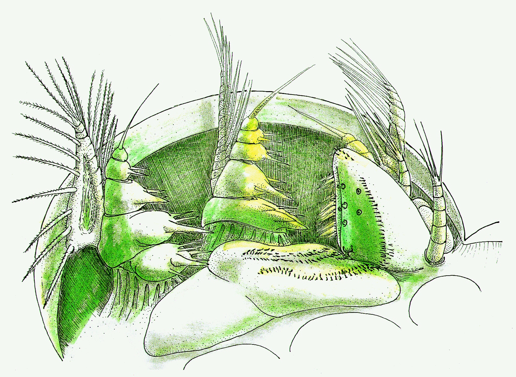
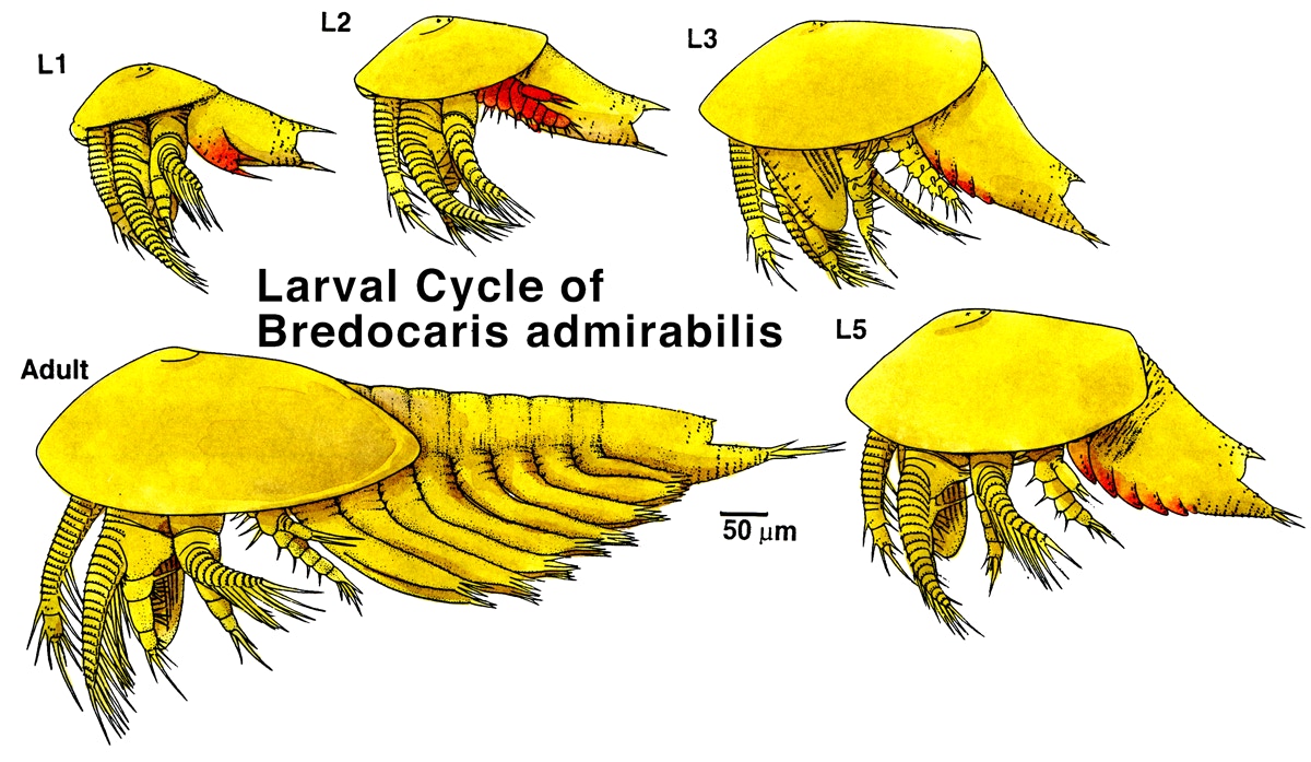
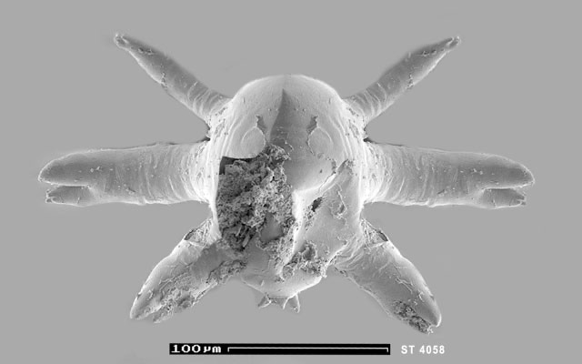
Below a few examples: set of size-adjusted drawings of crustaceans published until 1993; photographs arranged around the reconstruction of the ventral side of Agnostus pisiformis; generalized larval feeding apparatus of phosphatocopids; larval cycle of Bredocaris admirabilis; colourized sketch to help understanding the inner organization of Agnostus pisiformis.
4.2. 3D Reconstructions and 3D Modelling
Clay models
Already in 1987 Dieter started with 3D using modelling clay (plasticine) as a way to understand topologies and even movements in a better way and a method to improve reconstructive drawings. The first paper, in which this was presented was the work on Bredocaris admirabilis, published in 1988. The plasticine model was 40 cm large and built as a half model, with the appendages of one side left away. By this one could even apply macrophotography (e.g. of the mouth area) or produce serial images by progressively changing orientation of the limbs, so simulating movements. By combining tilted images, it was possible to produce complete views even from oblique angles.
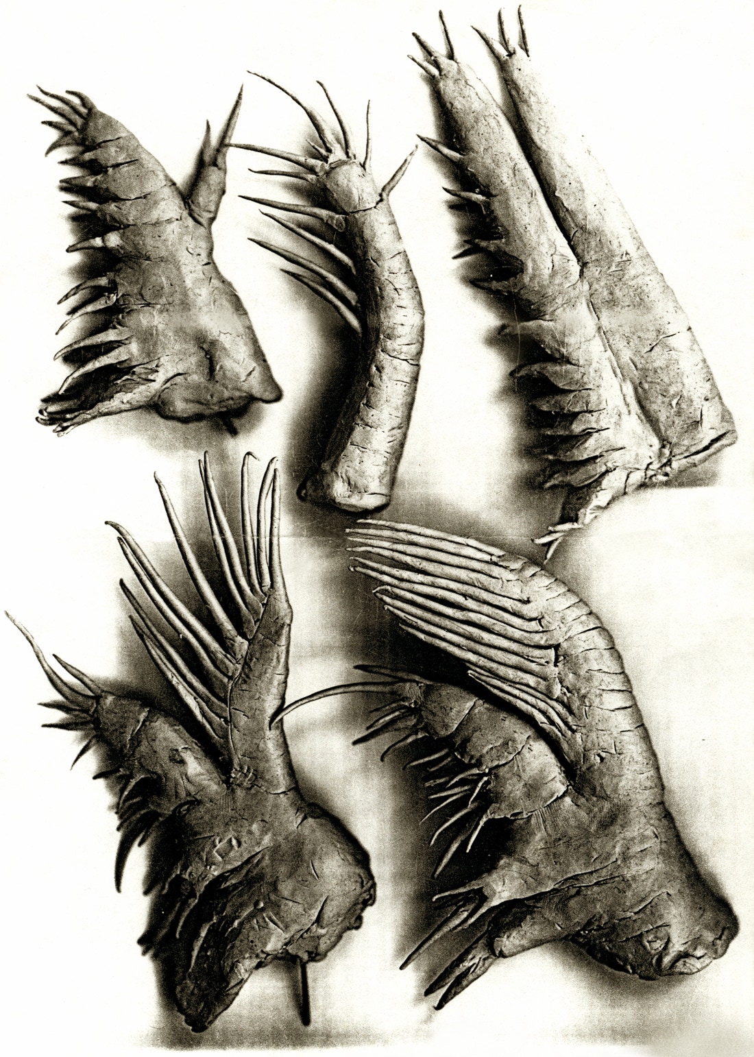
Left: Photograph of the model, right side mirrored; to the right the redrawing; plasticine models of the appendages, modelled from the pictures in the monograph.
We also experimented with short movies by photographing each tilting step under the SEM and then combined the images into a movie.
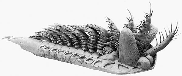
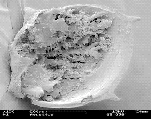
Left: a trunk shield of Agnostus pisiformis with appendages; right: pentastomid larva.
Combining photographs yielding more complete view of the animal
Another useful way to receive more complete views of 'Orsten' fossils was to combine photographs of different specimens, to e.g., multiply and re-scale appendages, or to mirror images. Examples are presented below:
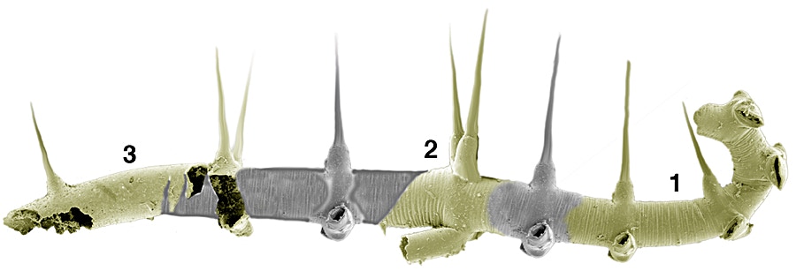
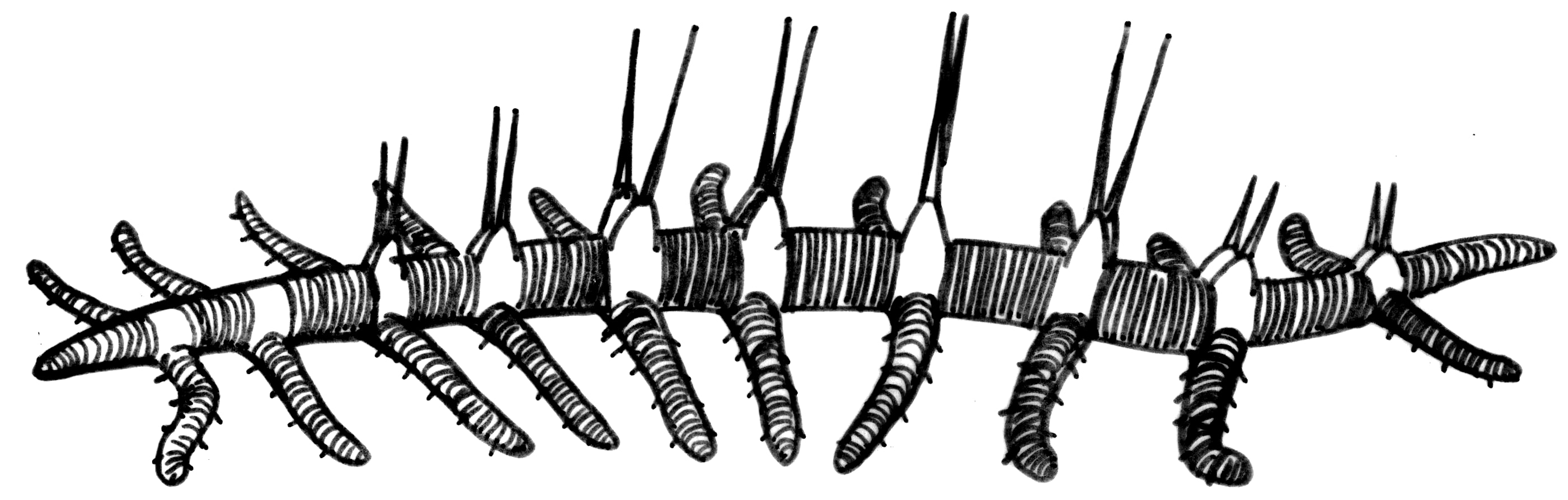
- ▪ Dala peilertae, with damaged head and rather complete thorax, and complete abdomen plus the furca added and an isolated limb of the right side serially added to fill up the set;
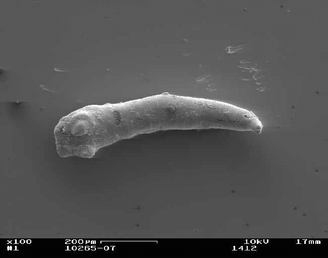
- ▪ the lobopodian Orstenotubulus evamuellerae (named in honour of the wife of Klaus, Eva), where the 3 specimens were combined by filling supposed gaps by the anterior segment of the right specimen; spines elongated in Photoshop; reconstruction underneath;
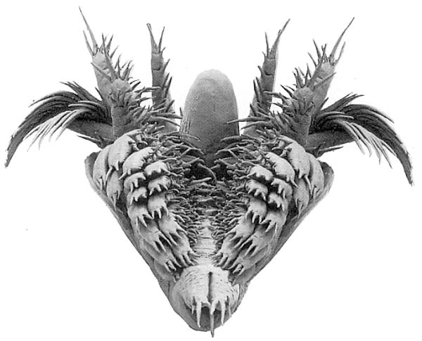
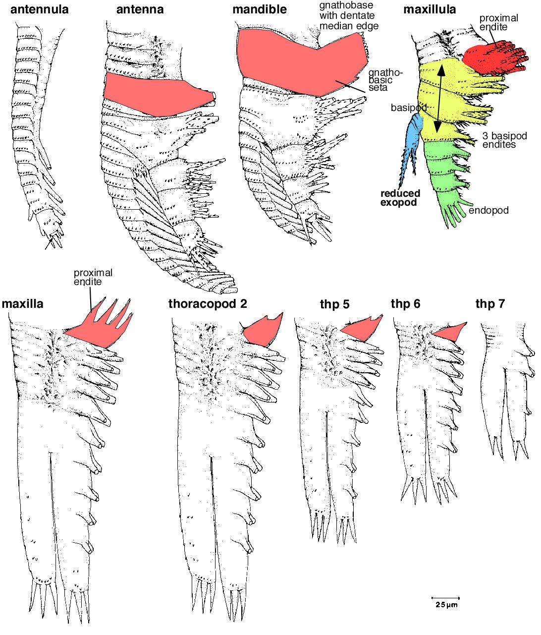
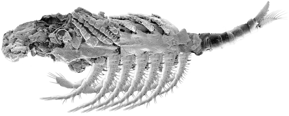
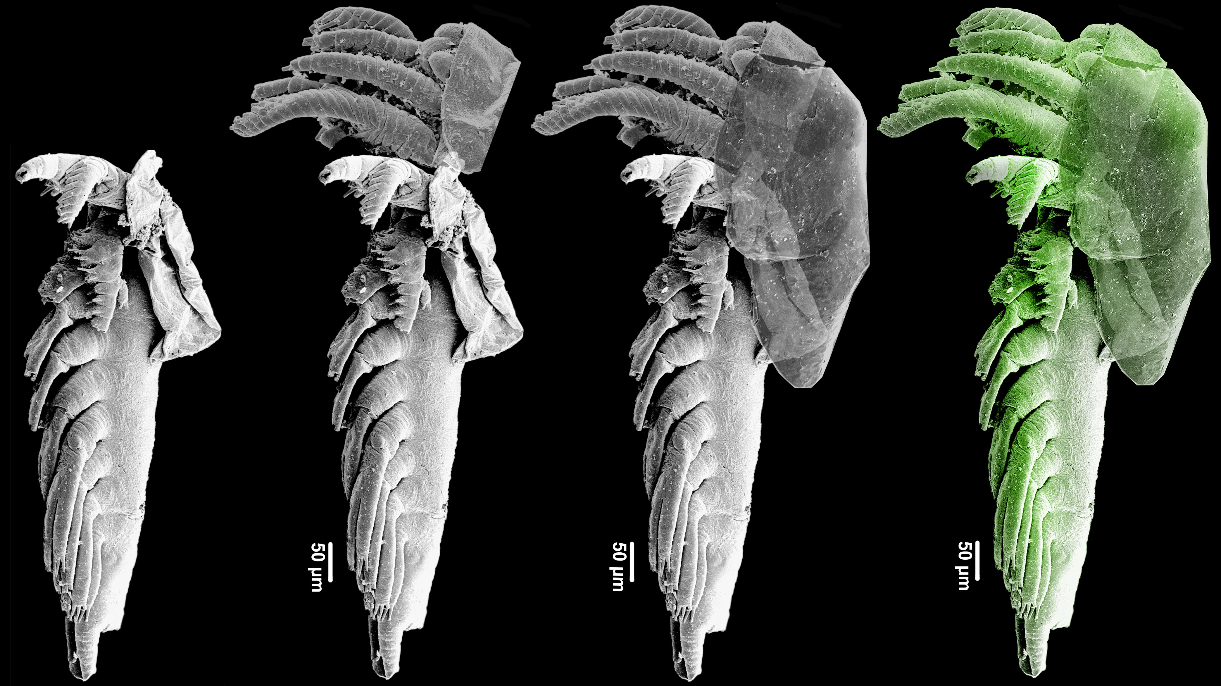
- ▪ "dissection" of appendages from SEM images of one or different specimens, which were later recombined to produce an as complete view of the animal – example Cambropachycope clarksoni;
- ▪ the second type of so-called type-A larvae, which differs in several aspects from the one originally described in 1986 by Klaus and Dieter (second type published in 1990); damaged right side substituted by the mirrored left side;
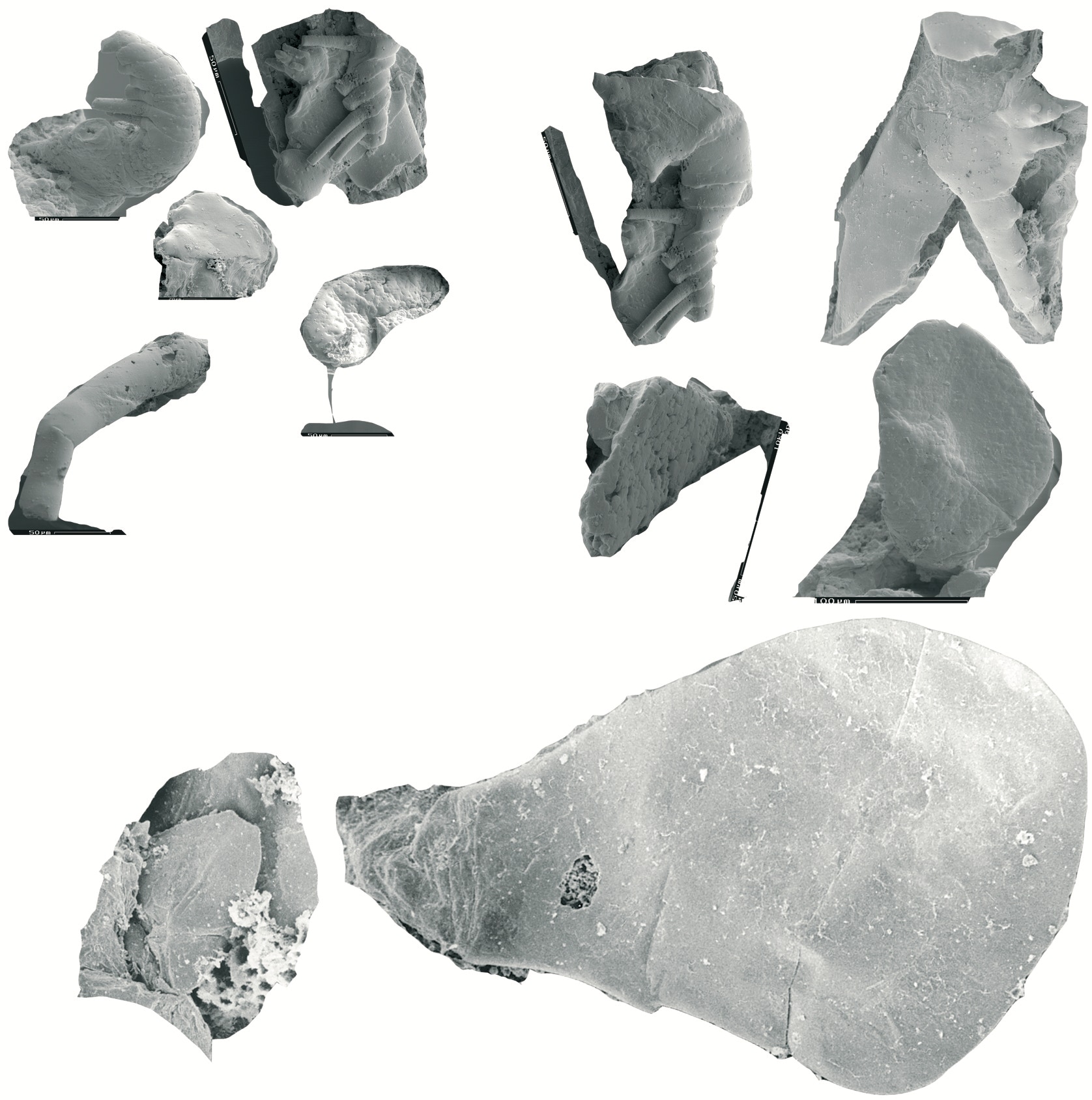
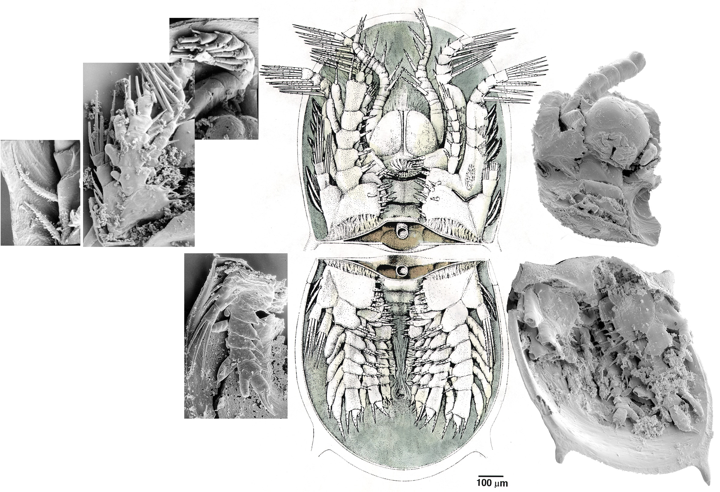
- ▪ a picture series of the holotype of Bredocaris admirabilis, with three fragments added to it: head part, shield and finally the colourized image.

© 2020 CORE
